We study how embryos with high potential differ in actively expressed genes.
Contact: Tereza Toralová
The aim of this research plan is to find out, which proteins are crucial for early stages of embryonic development. The obtained knowledge will help to further optimize culture conditions, to increase the rate of oocyte fertilization and improve the ability of the embryos to implant into the endometrium.
To describe the molecular processes connected to high developmental potential of the embryos, we will describe the translatome of high and low quality embryos and describe genes, whose translation is distinctive for high quality embryos. Translatome is a set of mRNAs that are currently bound to ribosomes and thus are being translated into proteins. At the beginning of the embryogenesis, the early embryos are transcriptionally inactive, and the regulation of embryonic development is driven at the post-transcriptional level. This means primarily the regulation of maternal mRNA translation, post-translational modifications and regulation of protein degradation. All the mRNAs are of maternal origin at that time, i.e., it was synthesized in oocyte and handed over to the arising embryo. The analysis of translatome is the most suitable indicator of the currently needed proteins in the cell. As the development proceeds, the inherited mRNAs and proteins are degraded, and the embryo takes over the control. Whilst maternal mRNAs are degraded in general, degradation of maternal proteins is much more specific, both concerning targeting to degradation (some proteins need to be degraded before initiation of embryonic transcription, some other are stored throughout the whole preimplantation development) and interspecies comparison. A similar specificity was found also in control of protein translation. Our aim is to describe the signalling pathways that are crucial for proper course of embryonic development and whose inadequate expression causes developmental arrest or low quality of the embryos.
The main goal is thus to reveal the cause of low developmental competence, optimize culture conditions and provide optimal environment for embryo culture. The obtained results will be discussed with the collaborating IVF clinics, so that the data are optimally utilized in human medicine.

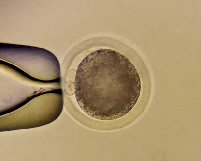
Figure1: Zygote (1-cell stage embryo) prepared for micromanipulation. (Veronika Kinterová)

Figure 2: Cumulus-oocyte complex with fluorescently labelled nucleoli in cumulus cells. (Tereza Toralová)

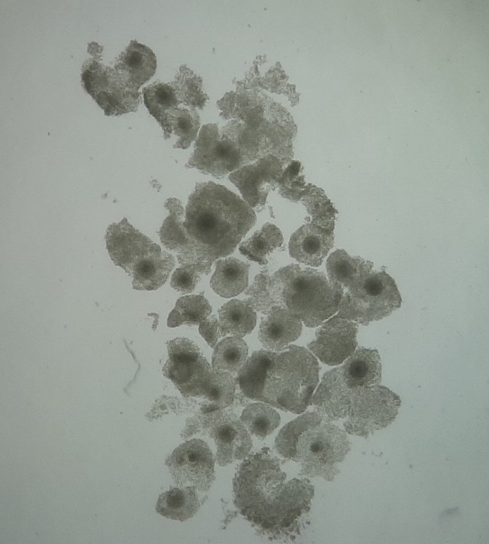
Figure 3: Bovine cumulus-oocyte complexes (Veronika Kinterová)

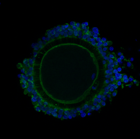
Figure 4: Transzonal projections in cumulus-oocyte complex (Veronika Kinterová)


