We investigate preimplantation differences between embryos with high and low developmental potential.
Contact: Tereza Toralová
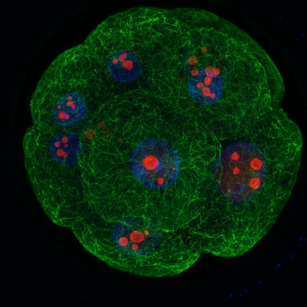
Our objective is to document early embryonic development of cattle, spanning from zygote to the blastocyst stage, the last stage before implantation into the endometrium. The obtained data will serve as a basis for artificial intelligence, which will “learn” to non-invasively recognize tiny differences in quality of individual embryos. This will enable to recognize embryos with the highest developmental potential.
Recently, a significant progress of artificial reproduction techniques has been made both in human medicine and in animal research model organisms. Nevertheless, many embryos do not dispose a high developmental potential. Therefore, the evaluation methods have to be improved. The aim of our project is to monitor preimplantation development of cattle using time-lapse imaging directly in the incubator without disturbing the culture conditions. The obtained videos will serve to develop AI-based tools to predict, which embryo have the highest chance to implant into the endometrium. Bovine embryos are the best model for such a research, as their development closely resembles human development, but are much more available than human embryos. The main output of this project will be detailed profile of normal preimplantation development of mammalian embryo, identification of critical moments of the development and optimization of culture methods for improvement of IVF successful rates. The results will not only be published in research journals but will be applied in practice and in education as well. The obtained data will be used as a basis for subsequent research plan of this project, translatome profiling, in which we focus on a so called translatome, an active set of genes that are currently used by the embryo. These data will help us to reveal key signalling pathways responsible for successful development.

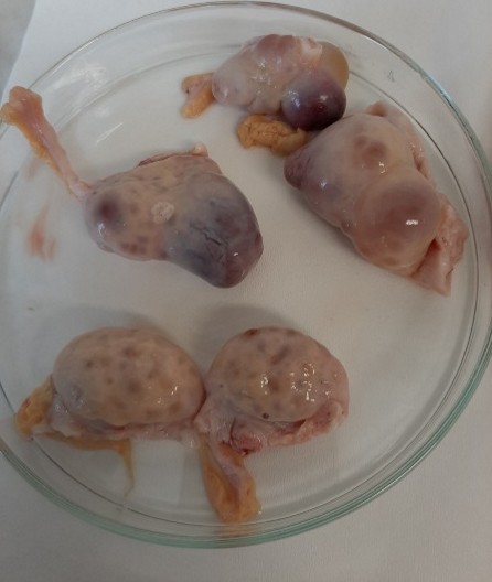
Figure 2: Bovine ovaries (Veronika Kinterová)

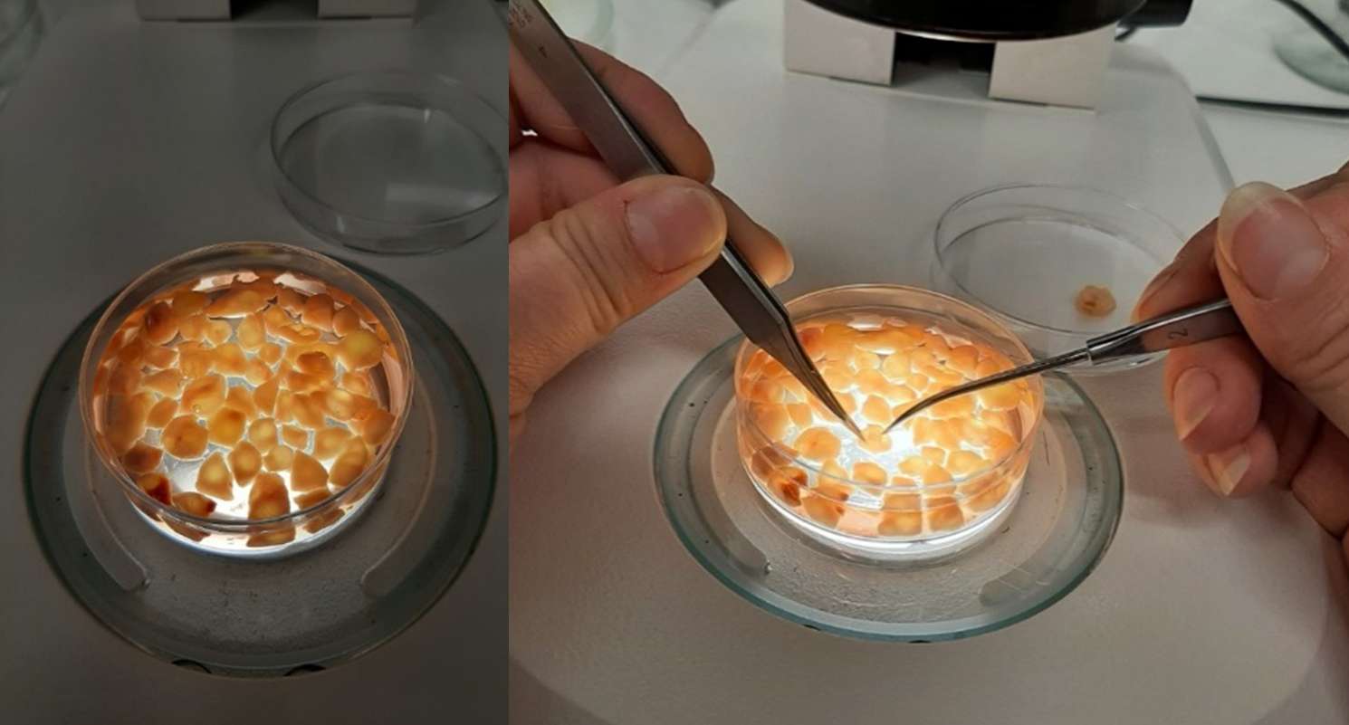
Figure 3: Isolation of bovine oocytes from dissected follicles. (Veronika Kinterová)


