Regulation of the cell cycle in the egg and early embryo – how cells ensure the proper onset of development.
Contact: Martin Anger
Early development in mammals is characterized by errors, which might lead into embryo loss. And the reason behind this situation might be the differences between cell cycle and chromosome segregation control mechanisms between somatic cells and germs cells or embryos. One of the biggest difference is a spectrum of molecules utilized by somatic cells for control of their cells cycle. Whereas somatic cells express various types of cyclins (A, B, D and E) and CDK kinases (CDK1 a CDK2), it seems that the embryos are using merely Cyclin B and CDK1 while other molecules are expressed later during the development.
Similar situation can be demonstrated in control mechanisms of chromosome segregation. Somatic cells utilize pathway called Spindle Assembly Checkpoint (SAC). This pathway ensures that all chromosomes within the cell are correctly attached to the spindle. And until this is achieved, the cell cycle progression is blocked. When the spindle is assembled correctly, and all chromosomes are engaged, the SAC is turned off, which leads into activation of large protein complex called Anaphase promoting complex/Cyclosome (APC/C). Activity of this complex than leads into segregation of chromosomes into daughter cells.
Our results as well as results from other laboratories showed, that oocytes are unable to precisely control the segregation of their chromosomes. And recently we have shown, that early embryos even do not utilize SAC pathway. It is obvious that these and other differences between cell cycle and chromosome segregation control might eventually lead into embryo loss.
To study the cell cycle and chromosome segregation in germ cells and early embryos we utilize several animal model organisms to obtain results relevant for all mammals, including human. We will use techniques allowing to express various biomolecules within oocytes end embryos, such as RNA. And for monitoring cell and chromosome division, we employ live cell imaging techniques.
The results will contribute to our understanding of the origin of errors in cell cycle and chromosome segregation control in germ cells and embryos. Simultaneously, we will focus on procedures allowing to correct such errors in order to prevent loss of germ cells or embryos.


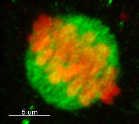
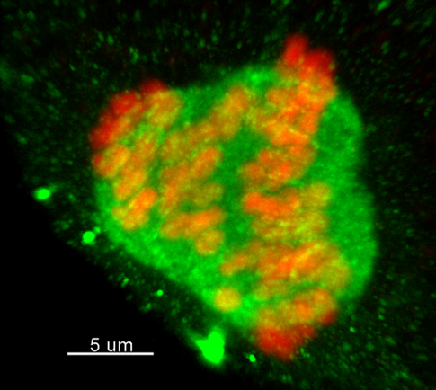
Figure 1: Normal (left) and tripolar (right) spindle in meiosis I in cattle oocytes. Chromosomes are in red, tubulin in green. Authors: Karolína Litošová, Martin Anger

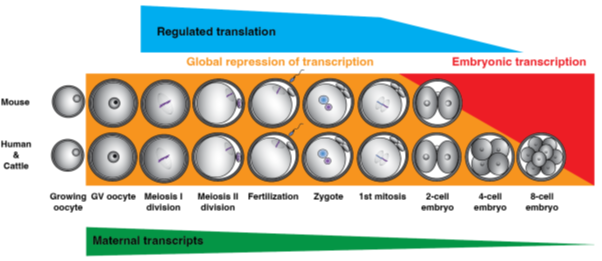
Figure 2: Obstacles and pitfalls of early embryonic development. The picture illustrates a period when genomic translation is silenced and complex events, such as the two meiotic division, fertilization and the first cleavage cycles of a newly developed embryo a controlled mostly by translation. Authors: Lenka Radoňová, Martin Anger


