
AI for Embryo Viability

We use deep learning and artificial intelligence to classify embryo development from time-lapse microscopy data.
Contact: Pavel Hozák
The development of a healthy embryo begins with the proper maturation of the egg (oocyte) and continues after fertilization. Understanding how eggs and embryos grow is crucial for fertility and for improving the success rates of assisted reproduction (ART), such as in vitro fertilization.
As part of the ART project, we focus on tools powered by artificial intelligence (AI) that automatically analyze how oocytes and embryos develop. By combining modern microscopy and AI, we can monitor embryos in real time, record important developmental features, and predict their quality.
We use gentle imaging techniques, such as time-lapse microscopy and light-sheet microscopy, which allow continuous observation of embryos without damaging them. By labeling specific cellular structures, we can track how oocytes mature and how embryos divide and grow up to the blastocyst stage.
Because human eggs and embryos are limited for research, we use animal models, such as mice and cattle. These models allow us to test new imaging methods, manipulate genes or metabolism, and create large datasets for training AI.
Our AI models focus on key developmental parameters, such as the timing of oocyte maturation and chromosome condensation, chromosome segregation during cell division, the size and position of the mitotic spindle, the duration of cell cycle phases, and the position of the nucleolus.
Using deep learning methods, we track cells in microscopy videos and classify their developmental stages. This approach allows us to combine data from different types of microscopy into a single AI model and improve its prediction accuracy. Once trained, AI models could automatically assess the quality of oocytes and embryos, helping doctors select the most promising embryos for transfer.
In short, by combining biology and AI, our work aims to achieve a better understanding of embryo development, improve embryo selection for assisted reproduction, and increase the success rates of procedures such as in vitro maturation and the use of cryopreserved oocytes and embryos.

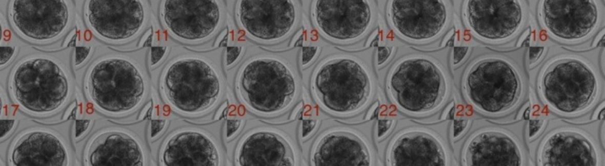
Figure 1: Timelapse monitoring of cattle embryo development. The sequence of images covers approximately 6 days of development. Authors: Tereza Pauerová, Martin Anger
Figure 2: Four cells cattle embryo. Authors: Tereza Pauerová, Martin Anger