


Programme
Takashi Akera, National Heart, Lung, and Blood Institute, USA
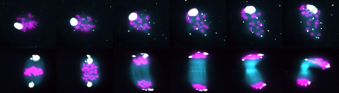
Mendel’s Law of Segregation states that each allele has an equal chance to transmit to the next generation. However, this law can be violated by selfish genetic elements, which manipulate the production of gametes (e.g., eggs, sperm) to increase their own transmission rate. This genetic cheating in meiosis, meiotic drive, has significant impacts on Genetics, Evolution, and Reproduction because the cheating alters transmission ratios and manipulates gametogenesis, often leading to fertility issues and genetic disorders (e.g., Down Syndrome). In female meiosis, selfish elements bias their transmission by preferentially segregating to the egg. However, it remains largely unknown how these elements bias their segregation to the egg especially in animals. My lab uses mouse models and cell biological approaches to visualize selfish elements to reveal how these elements manipulate the female germline to preferentially transmit to the next generation. I will discuss our recent findings on the mechanisms of meiotic drive and how it impacts mammalian reproduction and speciation.
Keynote lecture: Spatiotemporal regulation of the oocyte spindle: New insights into aneuploidy origins
Ahmed Balboula, University of Missouri, USA
Chromosome mis-segregation frequently occurs during oocyte meiosis I, leading to aneuploidy, the major genetic cause of infertility and congenital abnormalities. Why female meiosis is error-prone is largely unknown. In mouse oocytes, spindle positioning is a dynamic process. The spindle is assembled and positioned at the oocyte center until metaphase I. Later, the spindle undergoes a dramatic migration towards the cortex in a time-sensitive manner, a critical process for the highly asymmetric division and faithful chromosome segregation. We employed time-lapse super-resolution microscopy, laser ablation, optochemical tools, and genetic approaches to unveil a new model for meiotic spindle positioning and timely spindle migration.
Marie-Emilie Terret, Center for Interdisciplinary Research in Biology, College de France, France
Female meiosis generates a single large oocyte through asymmetric divisions controlled by cortical mechanics, yet their dysregulation is common in mammals and poorly understood. Here we show that increased cortical contractility drives spontaneous oocyte polarization mid-meiosis I. RhoA/ROCK-mediated activation of myosin-II induces a polarized cortical domain enriched in actomyosin, opposite the division site. This ectopic polarity generates directional cytoplasmic flows that accelerate and misorient spindle positioning, consistent with quantitative analysis and mathematical modelling. These flows reorganize the oocyte cytoplasm, disrupting organelle distribution and reducing their inheritance after meiosis I. In parallel, cortical granules and the sperm receptor Juno accumulate within the polarized domain, impairing fertilization and early embryonic development. Similar phenotypes are detected in mouse and human oocytes using machine learning, indicating that the mechanisms uncovered here are physiologically relevant. Together, our findings reveal that excessive contractility rewires oocyte polarity and cytoplasmic organization, with direct consequences for developmental potential.
Jerzy Dobrucki, Jagiellonian University in Kraków, Poland
DNA single-strand breaks (SSBs) occur in mammalian cells at thousands per day due to spontaneous loss of purine bases, oxidative damage, and errors in replication. They are repaired within minutes but pose a mutation risk if repaired improperly. Detecting and quantifying SSBs in tissue samples presents technical challenges and the numbers of SSBs at any given time in various mammalian tissues and cell types remains unknown. We detected SSBs in isolated oocytes, zygotes and cryosections of several murine tissue types using sSTRIDE technique. We found that oocytes had the lowest number of SSBs (below 1 in average) among all cell types examined. In somatic cells SSB numbers varied significantly not only between tissues (average numbers up to 10/cell) but also among individual cells within the tissue. Interestingly, high-metabolic-demand tissues, like the brain, skeletal and heart muscle, exhibited the highest levels of SSBs. These results provide the first detailed description of inter- and intra-cell variability of the SSB frequency in an organism and underscore the unique characteristics of female germ cells, which are known to have at least an order of magnitude lower frequency of de novo mutations than somatic cells.
Suzanne Madgwick, Newcastle University, United Kingdom
Human oocytes are highly prone to cell cycle errors that can result in cell cycle arrest, chromosome segregation errors during meiosis I, and ultimately cell death. Compared with mitotic cells, the mechanisms underlying these errors remain poorly understood. A major limiting factor in oocyte research is material availability, with low numbers of oocytes recovered from mammalian model organisms and human oocyte research being further hampered by significant and legitimate ethical considerations. Because of this material bottleneck, current biochemical analyses are limited. There is insufficient material for traditional mass spectrometry, which, even when feasible, lacks subcellular spatial information, while immunofluorescence-based imaging techniques can analyse only a small number of targets simultaneously. Consequently, our understanding of the spatiotemporal regulation of key cell cycle events in mammalian oocytes remains limited. To address this shortfall, we have developed a protocol to apply Imaging Mass Cytometry (IMC) to mammalian oocytes. IMC utilises heavy-metal-conjugated antibodies and sequential laser ablation to generate 2D images with 1 μm² pixel resolution across up to 40 channels simultaneously. Our current panel of 32 antibodies allows us to begin constructing a highly multidimensional model of the oocyte cell-cycle proteome throughout meiosis, capturing protein abundance, localisation and phosphorylation states. Here, we present results demonstrating the power of IMC to identify proteome-level changes during meiotic progression and maternal ageing, enabling the identification of likely non-competent oocytes.
Lukas Ded, Institute of Biotechnology of the Czech Academy of Sciences, BIOCEV, Czech Republic
Fertilization is highly complex process which, in mammals, includes intricate interaction of two gametes - sperm and oocyte, leading to the creation of the zygote and subsequent development of the new organism. The gamete interaction starts with the contact of the sperm with the cumulus oophorus, and continues with contact and penetration of the zona pellucida, passing through perivitelline space, contact and fusion with oolemma and further steps inside the ooplasm. All individual steps takes place in highly intricate 3D environment and are facilitated by the interaction of complex cellular, sub-cellular and molecular parts of the gametes. All these complex aspects make the fertilization process a challenge to be properly imagined and rigorously visualized. The aim of our project is to combine the physical, microscopic and numeric data with 3D modelling to create visually educative as well as physiologically relevant 3D models of the fertilization process integrating its cellular, sub-cellular and molecular aspects. Such models will further improve our understanding of the process of gamete interaction and fertilization and enable to develop new concepts and hypotheses throughout its individual steps.
Michaela Frolikova, Institute of Biotechnology of the Czech Academy of Sciences, BIOCEV, Czech Republic
Juno and CD9 are two essential proteins of the mammalian oocyte plasma membrane, yet their functional relationship has remained unclear. This presentation summarizes evidence demonstrating their spatial organization within distinct oolemma compartments and their close association in the microvillar region, providing new insight into the molecular organization of the oocyte surface before fertilization.
Helena Fulkova, Institute of Experimental Medicine of the Czech Academy of Sciences, Czech Republic
Female reproductive ageing is accompanied by a progressive decline in oocyte quality, increased chromosomal abnormalities, and reduced developmental competence. Accumulation of DNA damage in fully grown oocytes has been considered a major contributor to this process and is generally viewed as irreversible. In our study, we tested whether age-associated DNA damage can be reversed by exposing chromosomes from advanced maternal age (AMA) oocytes to the nuclear environment of young oocytes. Using a series of germinal vesicle (GV) transfer and selective enucleation experiments in the mouse, we demonstrated that AMA oocytes exhibit markedly elevated levels of DNA damage, as indicated by γH2AX-positive chromosomal foci, together with increased rates of chromosome segregation errors. Surprisingly, transfer of aged GVs into cytoplasts enriched with the soluble karyoplasmic fraction of young oocytes substantially reduced DNA damage signals and decreased the frequency of chromosomal abnormalities. In contrast, conventional GV transfer or replacement of the oocyte nucleolus alone failed to produce a comparable effect, indicating that DNA repair competence resides within soluble, non-chromatin-bound nuclear factors rather than the nucleolus itself. Functional analyses further revealed partial restoration of chromatin dynamics in reconstructed oocytes, consistent with improved accessibility of damaged chromatin to repair mechanisms. Most importantly, these molecular and cytogenetic improvements translated into enhanced developmental competence, with reconstructed AMA oocytes supporting full-term development, whereas intact aged oocytes failed to produce offspring. Together, our findings demonstrate that age-dependent DNA damage in oocytes is not necessarily permanent and can be substantially mitigated by factors present in the karyoplasm of young oocytes. These results redefine current views of oocyte ageing and establish a proof-of-principle for cell-based strategies aimed at rejuvenating female gametes and restoring reproductive potential.
Rebeca Collier, University of South Bohemia, Czech Republic
In mammalian preimplantation embryos, different cell lineages occupy specific niches. For example, the outer trophectoderm (TE) comprises a monolayer of epithelialized cells surrounding the inner-cell mass (ICM) and blastocyst cavity. In mice, TEAD4 is known as a transcription factor that regulates TE-specific genes in a polarity-dependent manner during TE specification. Here we show that it also maintains blastocyst TE integrity, as knocking down (KD) Tead4 via clonal siRNA causes abnormal morphology of outer-cell apical domains, which correlates with the atypical contribution of Tead4-KD cell clones to an enlarged ICM throughout blastocyst maturation; with only minimal feedback on established apical polarity. Light-sheet live-cell embryo imaging reveals these cells either actively migrate into the ICM, sometimes involving apical domain abscission, or are positioned post-division, linking disrupted apical morphology to cell repositioning. RNA-Seq data indicate TEAD4 regulates genes related to the cytoskeleton, particularly actin, and cell adhesion, which we propose are required for the appropriate maintenance of the spatial positioning of specified TE cells in the blastocyst. Indeed, knocking down Tead4 in combination with two identified target genes, the atypical GTPases Rnd1 and Rnd3, partially rescues aberrant outer-to-inner cell allocations but does not influence the onset of apical domain morphological abnormalities. These findings indicate that Tead4 and its regulated transcriptome actively contribute to the maintenance of the outer TE lineage until the peri-implantation stage.
Emmanuel García Sánchez, Institute of Animal Physiology and Genetics of the Czech Academy of Sciences, Czech Republic
In oocytes, mRNA synthesis is regulated by the TBPL2/ RNA Pol II machinery. Transcriptome long-term stability relies on membrane-less structures associated with the cytoskeleton termed CytoPlasmic Lattices (CPLs) and on poly(A) shortening. Tbpl2-/- oocytes stop growing. Could this growth arrest be linked to defects in transcript stability control? My research shows that the mRNA-associated stability proteins are indeed expressed during oocyte growth and that their expression and localization are altered in the Tbpl2 mutant. Histological and subcellular analyses show cytoplasmic anomalies and expanded nuclear foci where poly(A) accumulates. Electron microscopy of the Tbpl2 mutant highlights mitochondrial and golgi clustering near the nucleus and a complete absence of CPLs formation. These findings emphasize the critical role of oocyte growth in regulating maternal transcriptome stability.
Kianoush Kakavand, Institute of Animal Physiology and Genetics of the Czech Academy of Sciences, Czech Republic
Translational control is a fundamental regulatory mechanism during mammalian oocyte development, particularly throughout meiotic maturation, when transcription is globally silenced. Consequently, precise temporal regulation of protein synthesis is essential for coordinating chromosome segregation, cytokinesis, and the acquisition of developmental competence. Here, we demonstrate that global protein synthesis is transiently suppressed at defined stages of meiotic progression in mouse oocytes. We further show that this stage-specific translational repression functions as a critical regulatory mechanism that promotes meiotic progression and successful oocyte maturation. Collectively, our findings establish dynamic translational regulation as a key determinant of terminal oocyte development.
Abstracts of the lectures, and the final program, including a full list of speakers, will be updated and made available on this website.